2012.11.22
Setting up the Museum of Microscopy
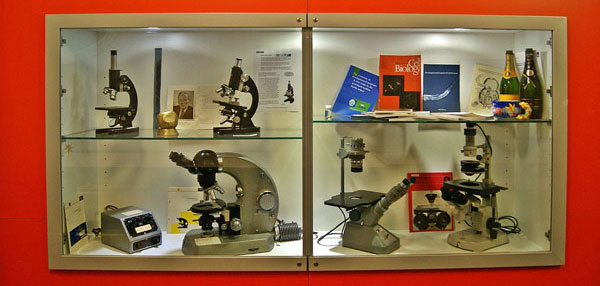
Beck Kassel CBS; Olympus; Zeiss
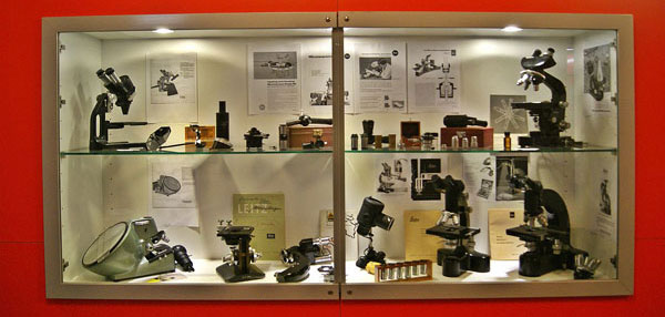
Leica Window (Reichert; Wild Heerbrugg; Leitz)
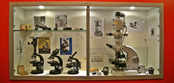
Carl Zeiss Window
Cell Biology Utrecht University, 5th floor… Read more
2012.11.22
Setting up the Museum of Microscopy

Beck Kassel CBS; Olympus; Zeiss

Leica Window (Reichert; Wild Heerbrugg; Leitz)

Carl Zeiss Window
Cell Biology Utrecht University, 5th floor… Read more
2012.11.20
Testing AMG EVOS digital inverted microscope
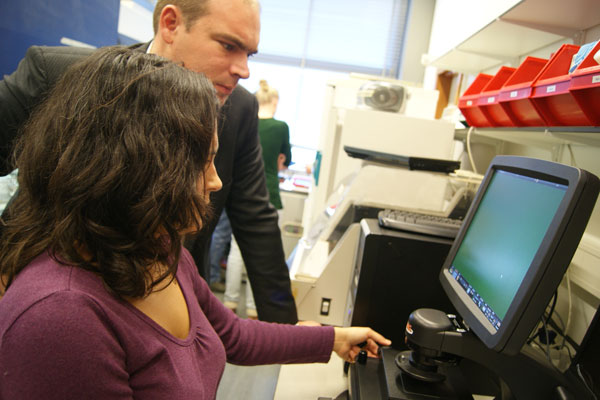
Sabrina Oliveira is testing 14C head and neck cancer cells incubated with
anti-EGFP nanobody, conjugated with far-red 800CW fluorophore from Licor
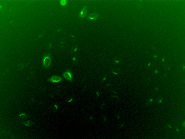
HeLa Rab6-GFP stable line, x20 objective
Cell Biology Utrecht University, Room N-527… Read more
2012.11.13
Testing the FLoid® Cell Imaging Station (Life Technologies / Invitrogen)
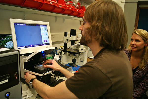
Benjamin Bouchet is checking the colony of the primary mammary cells
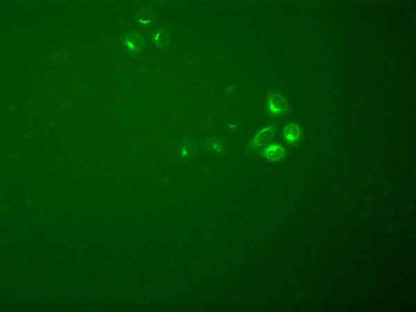
HeLa Rab6-GFP stable line
Cell Biology Utrecht University, Room N-527
2012.11.07
Testing the confocal microscope Leica TCS SP8 at Hubrecht Institute for Developmental Biology and Stem Cell Research
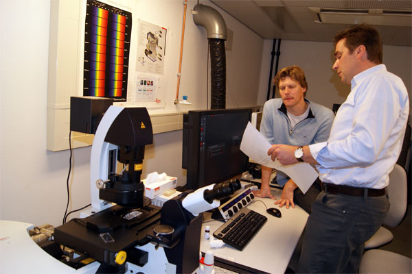
Marco Meijering (Leica Microsystems CMS) is explaining to Dr. Lukas Kapitein the advantages of the hybrid detection of the Leica TSC SP8 … Read more
2012.09.04
Testing Tokai Hit high grade controller
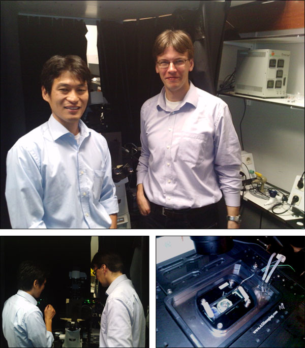
Toru Yamaguchi (Tokai Hit ) and PhD student Robert van den Berg
are discussing the temperature sensor of the improved
Tokai Hit stage top cell incubator.
Cell Biology Utrecht University, Room Z-514
2012.08.31
Testing the stereomicroscopes
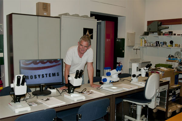
Frits Kindt after testing the stereomicroscopes
Leica EZ4HD; Leica EZ4; Zeiss Stemi DV4; Nikon SMZ445.
Cell Biology Utrecht University, Room O-523… Read more
2012.08.22
Testing of the super-resolution microscope Leica TCS SP8 STED at Leica Microsystems CMS GmbH (Mannheim)
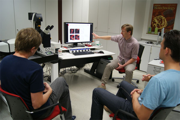
Ulf Schwarz (Leica Microsystems CMS) explains the working principles of gSTED.

confocal gSTED
Fixed microtubules… Read more