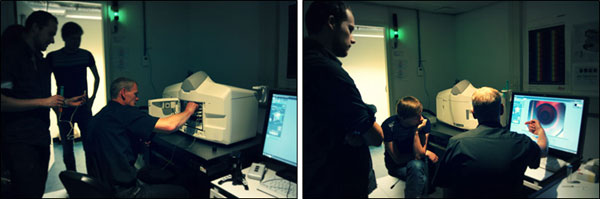
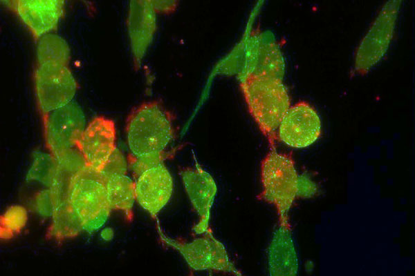
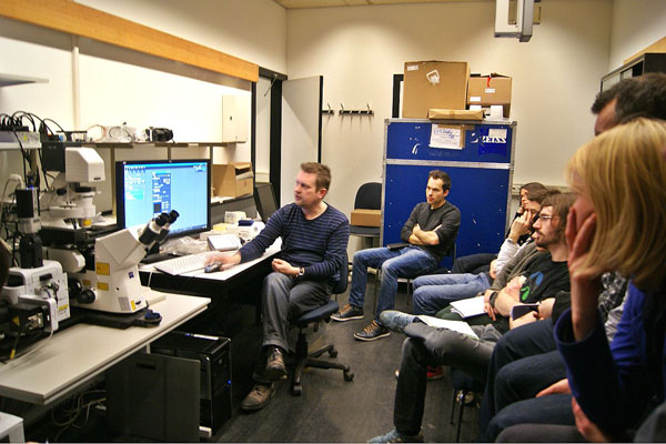
Utrecht Microscopy Summer Meeting Focus on: Image Analysis Software
2013.09.03
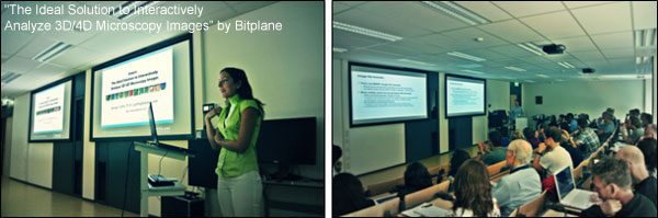
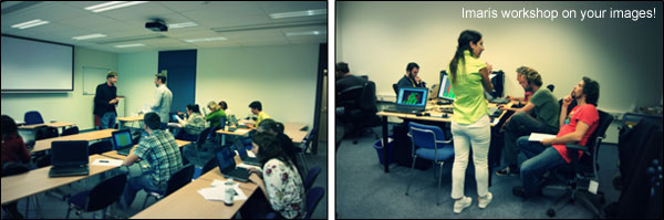
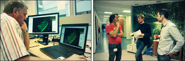
Utrecht Microscopy Summer Meeting Focus on: Image Analysis Software Huygens, Volocity, Imaris, ImageJ
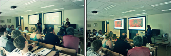
Vondelzaal, Stratenum building, UMC Utrecht
Link to the program: http://www.bioimaging-utrecht.nl/meetings/2013-seasonal-utrecht-microscopy-meeting/