BIC
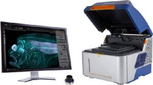
BioSPX Cytation C10& BioSPA 8 demo
2023.02.06-13
BioSPX Cytation C10& BioSPA 8 demo
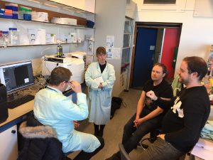
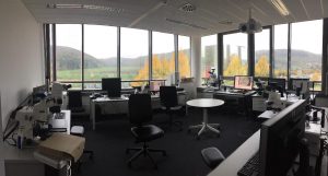
Cell Biology Utrecht University, Room O.512
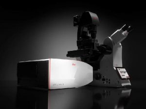
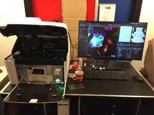
Testing Zeiss Elyra 7 and LSM 980 Airyscan2
2022.10.12-14
Testing Zeiss Elyra 7 and LSM 980 Airyscan2
ZEISS Microscopy Customer Center Europe, Oberkochen, Germany… Read more
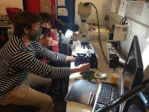
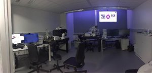
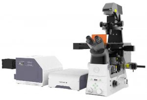
Nikon Ti2 with Spinning Disc Yokogawa SoRa W1 test
2022.08.01-16
Nikon Ti2 with Spinning Disc Yokogawa SoRa W1 test
Cell Biology Utrecht University, Room O.512… Read more
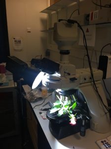
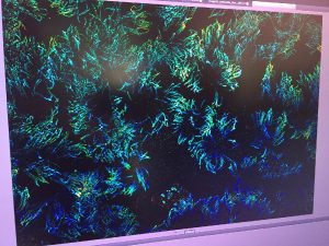
Zeiss Elyra 7 with Lattice SIM2 test
2022.04.07-15
Zeiss Elyra 7 with Lattice SIM2 test
Cell Biology Utrecht University, Room O.512… Read more